

Spine Signals: Your spine has been speaking, we are now listening!
This limitation has meant that, while the brain is widely studied, we know far less about how the spinal cord behaves in real time, especially during movement or in neurological diseases. When conditions such as Multiple Sclerosis (MS) or Motor Neuron Disease disrupt the signals passing through it, everyday actions gradually become harder. Yet researchers have had no standard tool to measure these changes.
Researchers in the NeuroMotor Group at Trinity College Dublin are working to change this. They have developed the SC10‑X/U system, a new way of recording spinal cord activity that could open doors to discoveries previously out of reach.
 regions") SC10-X/U Electrode System Design
SC10-X/U Electrode System Design
SC10-X/U is inspired by an internationally accepted EEG 10-10 system, which is a high-resolution method that standardises the placement of electrodes on the scalp. The newly-developed system applies similar principles—using specific, identifiable points as reference for consistent placement and uniformly-spaced electrodes and adapts them to the structure and shape of the spinal cord.
The development defines up to 76 standardised electrode positions, using anatomical landmarks such as the head, spine, and shoulder blades. This shared map allows for reliable and repeatable placement across participants and recording sessions.
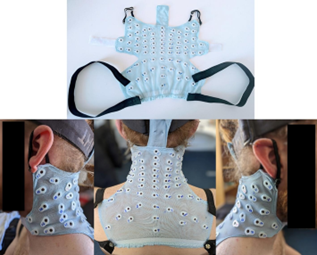
Figure 1 (left): Spinal cord model of the cervical and thoracic (neck and upper back) regions
 High-density Electrospinography (HD-ESG) Patch Implementation
The electrodes are embedded in a flexible and breathable patch that sits comfortably around the neck and upper back. The patch is:
Importantly, the system remains compatible with previous low-density configurations, enabling novel recordings to be compared with existing spinal cord research and reducing barriers for adoption.
Figure 2 (right): 1) Custom-developed electrode patch based on the SC10-X/U electrode system; 2) The electrode patch (up to 76 electrode channels) is placed on the skin over the neck and upper back area of the spinal cord (cervical and upper thoracic spinal cord).
Research Applications
The SC10-X/U system is currently being used in MS research. Damage to the cervical spinal cord is closely associated with disability progression in MS, yet structural imaging alone does not always explain changes in symptoms over time. Two people with similar‑looking scans can experience very different levels of functional decline. By directly assessing how the spinal cord functions, rather than just its structure, researchers hope to discover new insights into why and how symptoms worsen. Besides MS, the system shows promise in studying motor neuron disease, stroke, spinal cord injury, and cervical dystonia conditions, where the spinal cord communication is often disrupted and yet remains understudied!
Validation for Future Applications in Clinical Research
To validate the method, recordings were collected from ten healthy adults using a 64-electrode configuration during a gentle median nerve stimulation at the right wrist. After removing physiological and environmental noise, clear spinal cord responses were detected. Two well-characterised signals, known as N13 and P9, appeared at expected time points following the stimulation. Mapping these responses showed that N13 activity was strongest over the mid-cervical region (C5-C7), confirming the reliability of the system to capture spinal cord activity with appropriate temporal precision, while also enabling spatial precision—a capability previously unavailable with low-density recordings.
Opportunities for Global Collaboration and Research Partnerships
Since the first presentation of the high-density ESG system developed in TCD, research groups across Europe have expressed interest in adopting this standardised system. The team is now looking for industry partners to create research‑grade patches suitable for large international projects.
Similar to how the EEG system transformed brain research, standardised and high-density spinal recordings have the potential to reshape how spinal cord function is studied. It marks a shift in research by moving beyond the view of the spinal cord simply as a passive pathway, toward recognising it as a dynamic, active contributor to movement and sensation.
Get Involved
Interested in SC10-X/U for your research? We are actively seeking industry partners and research collaborators to advance standardised spinal cord electrophysiology globally.
Contact: pmehra@tcd.ie
Learn More:https://futureneurocentre.ie/new-electrode-system-offers-a-window-into-spinal-cord-function/
Read the Full Publication: https://doi.org/10.1016/j.clinph.2025.2110824
 Dr. Prabhav Mehra | Postdoctoral Researcher
Prabhav is a postdoctoral researcher. He completed his PhD in the NeuroMotor research team, and has a master’s degree in Biomedical Sciences and a bachelor’s degree in Electrical and Electronics. His project focuses on investigating brain-body connections. His research interests include Neuromuscular Engineering, Signal Processing, Biomedical System Design, Model-based Biomarker, and Neurophysiology.
 Dr. Bahman Nasseroleslami | Group Leader, NeuroMotor Group / Principal Investigator / Associate Professor
Dr. Nasseroleslami received the B.Sc. degree in mechanical engineering from Iran University of Science and Technology, Tehran, Iran, in 2003, and the M.Sc. degree in biomechanical engineering from Sharif University of Technology, Tehran, Iran, in 2006. He received his Ph.D. degree in biomedical engineering from University of Strathclyde, Glasgow, Scotland, UK, in 2013. He was with the Department of Biology, Northeastern University, Boston, MA, USA, as a Research Associate (2012) and a Postdoctoral Research Associate (2013-2014). He joined the Academic Unit of Neurology in Trinity College Dublin, the University of Dublin, Dublin, Ireland as a Research Fellow in 2014, and then worked as a Senior Research Fellow. He is currently the Fr Tony Coote Associate Professor in Neuroelectric Signal Analysis in MND, a Principal Investigator, and the Research Strand Leader of the NeuroMotor research group at Trinity College Dublin.
- Article written by Lê Hoàng Vân Anh: Van Anh is currently an undergraduate Psychology student at Eötvös Loránd University, Hungary. She spent one semester as an exchange student at Trinity College Dublin, where she also volunteered with the NeuroMotor Group. Her main research interests lie in clinical and translational neuroscience, with side interest in cognitive neuroscience.
- Editorial support by Dr Sarah Cullen
Figures 1 and 2 https://ars.els-cdn.com/content/image/1-s2.0-S1388245725006765-gr1_lrg.jpg (Source: Mehra et al., 2025.
Retrieved from https://doi.org/10.1016/j.clinph.2025.2110824)